
Dutch study finds nuclear imaging accurately measures drug uptake in children with diffuse intrinsic pontine glioma In a first-ever molecular drug-imaging study in children, researchers in The Netherlands used whole-body positron emission tomography/computed tomography (PET/CT) scans to determine whether bevacizumab (Avastin) treatment of diffuse intrinsic pontine glioma (DIPG) in children is likely to be effective.
“Children with DIPG have a very poor prognosis, with less than 10
percent of the patients surviving two years from diagnosis,” explained Guus A. van Dongen, Ph.D., of VU University, Medical Center, Amsterdam, The Netherlands. “These tumors are resistant to all kinds of therapies. Chemotherapy, as well as new targeted therapies, may not reach the tumor due to the location within the brainstem.” For the study, researchers investigated whether bevacizumab can reach the tumor in children with DIPG by measuring the tumor uptake of zirconium-89 (Zr-89)-labeled bevacizumab with PET.
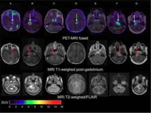
In addition, they evaluated the safety of the procedure and determined the optimal timing of imaging. Two weeks after completing radiotherapy, seven patients (age range: 6-17) were given whole-body PET/CT scans performed at 1, 72 and 144 hours post-injection. The optimal moment of scanning was found to be 144 hours post-injection. The patients also underwent contrast (gadolinium)-enhanced magnetic resonance imaging (MRI). “The results showed that indeed there is considerable heterogeneity in uptake of Zr-89-labeled bevacizumab among patients and within tumors,” Van Dongen pointed out. “This non-invasive in vivo quantification of drug distribution and tumor uptake may help to predict therapeutic potential, as well as toxicity, and could help develop strategies for improving drug delivery to tumors.” Van Dongen added, “Children with brain tumors and other solid cancers are particularly likely to benefit from molecular drug imaging, as drugs without therapeutic effect — based on a lack of drug-uptake in the tumor — may cause life-long side effects. Molecular drug imaging will open avenues for administering the right drug to the right patient at the most appropriate stage of the disease.”
For more information: www.jnm.snmjournals.org